ART Database
CLADE: H-Y-E (ARTC000002)
FAMILY: Diphtheria_C (ARTF000014)
Description
A family present in bacteria and viruses, it includes diphtheria toxin secreted by lysogenic strains of Corynebacterium diphtheriae.
InterPro - IPR022406
Diphtheria toxin (EC) is a 58kDa protein secreted by lysogenic strains of Corynebacterium diphtheriae. The toxin causes the disease diphtheria in humans by gaining entry into the cell cytoplasm and inhibiting protein synthesis [PUBMED:8573568]. The mechanism of inhibition involves transfer of the ADP-ribose group of NAD to elongation factor-2 (EF-2), rendering EF-2 inactive. The catalysed reaction is as follows: NAD + + peptide diphthamide = nicotinamide + peptide N-(ADP-D-ribosyl)diphthamide. The crystal structure of the diphtheria toxin homodimer has been determined to 2.5A resolution [PUBMED:1589020]. The structure reveals a Y-shaped molecule of 3 domains, a catalytic domain (fragment A), whose fold is of the alpha + beta type; a transmembrane (TM) domain, which consists of 9 alpha-helices, 2 pairs of which may participate in pH-triggered membrane insertion and translocation; and a receptor-binding domain, which forms a flattened beta-barrel with a jelly-roll-like topology [PUBMED:1589020]. The TM- and receptor binding-domains together constitute fragment B. This entry represents the N-terminal catalytic domain (also known as the C domain). This domain has an unusual beta+alpha fold [PUBMED:9012663]. The C domain blocks protein synthesis by transfer of ADP-ribose from NAD to a diphthamide residue of EF-2 [PUBMED:7833808, PUBMED:8573568].
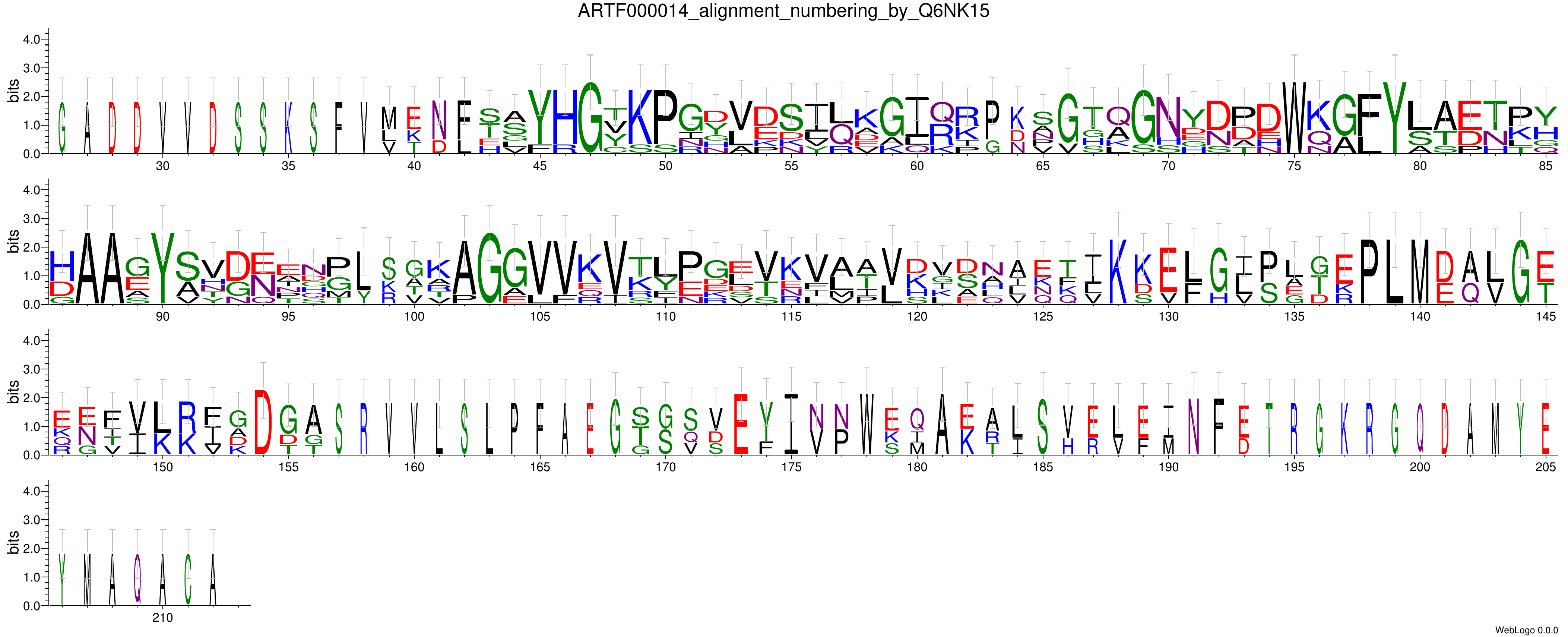
WebLogo

Sequences
Aligned domain sequences
Download sequenceUnaligned domain sequences
Download sequenceFull sequences
Download sequenceHMM Model
Structures
| Name | Structure | Additional informations |
|---|---|---|
| 1DDT | Download structure | Reference structure |
| 1DTP | Download structure | Reference structure |
| 1F0L | Download structure | Reference structure |
| 1MDT | Download structure | Reference structure |
| 1SGK | Download structure | Reference structure |
| 1TOX | Download structure | Reference structure |
| 1XDT | Download structure | Reference structure |
| 4AE1 | Download structure | Reference structure |
| 4AE0 | Download structure | Reference structure |
| 5I82 | Download structure | Reference structure |
| 7K7B | Download structure | Reference structure |
| 7K7C | Download structure | Reference structure |
| 7K7D | Download structure | Reference structure |
| 7K7E | Download structure | Reference structure |
Origin
Source: Pfam 34.0, rp75 Number of sequences: 6 Average length of the domain: 127 aa HMM: Model length: 147 Clustering level: 80% Alignment: ClustalO Additional information: sequences longer than 50 amino acids